Here’s a detailed overview of an X-ray of the knee—what it is, why it’s done, and what it shows:
🦴 What a Knee X-Ray Is
- An X-ray is an imaging technique that uses a small amount of radiation to create pictures of bones.
- A knee X-ray shows the bones, joint space, and alignment of the knee.
- Commonly used to diagnose injuries, arthritis, or other bone conditions.
🔍 Why a Knee X-Ray Is Done
- Fractures or Broken Bones – after trauma or injury.
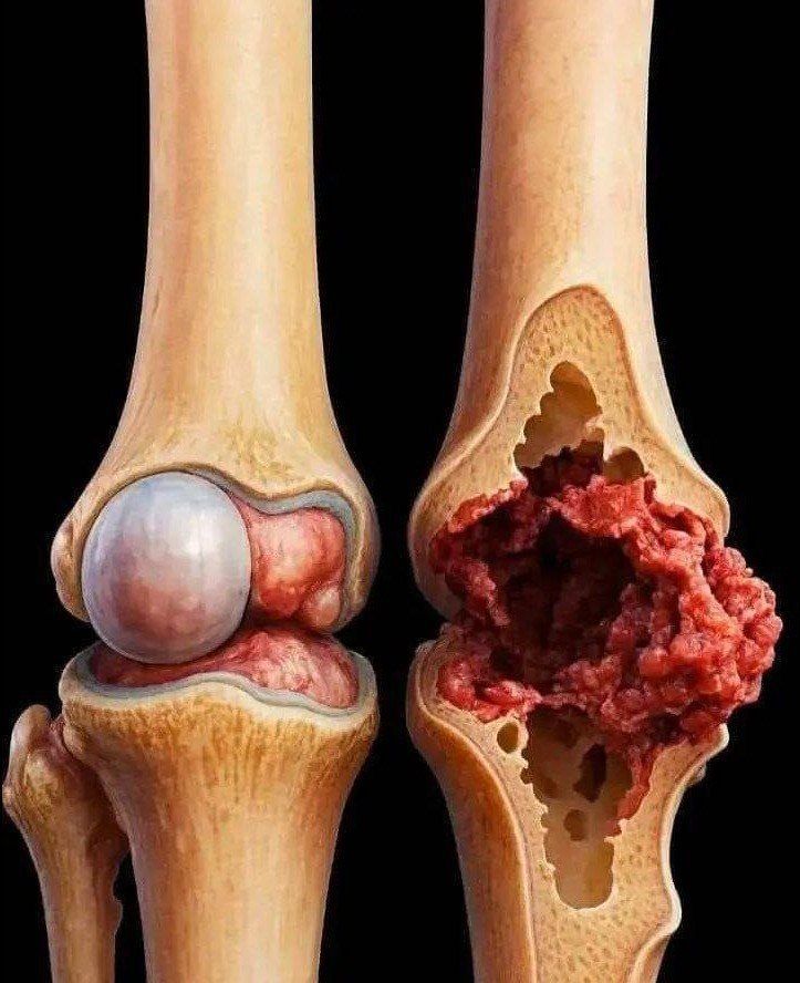
- Arthritis – to check for joint space narrowing or bone spurs.
- Bone Infections (Osteomyelitis) – to detect early changes.
- Knee Pain or Swelling – helps rule out structural problems.
- Pre-Surgical Assessment – before knee replacement or other surgery.
🖼️ Types of Knee X-Rays
- Anteroposterior (AP) view: Front-to-back image of the knee.
- Lateral view: Side view, shows joint alignment and soft tissue indirectly.
- Sunrise or skyline view: Focuses on the kneecap (patella).
- Weight-bearing view: Patient stands on the leg to assess joint under pressure.
✅ What Doctors Look For
- Bone alignment and fractures
- Joint space narrowing (arthritis)
- Bone spurs or osteophytes
- Signs of infection or tumors
- Degenerative changes
⚠️ Safety Notes
- X-rays use low-dose radiation, considered safe for most people.
- Inform your doctor if you’re pregnant, as extra precautions may be needed.
💡 Tip:
Sometimes X-rays are combined with MRI or CT scans if soft tissue (ligaments, cartilage, tendons) needs to be evaluated, since X-rays mainly show bone.
If you want, I can explain how to read a knee X-ray at home for common issues like arthritis or fractures—just a basic guide for non-doctors.
